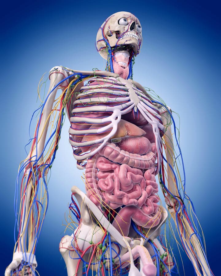
Abdominal Anatomy - Anatomy Of Human Abdominal Vein System Bath Towel for Sale by Stocktrek Images. The anterior abdominal wall (figs. Divided into 9 regions by two vertical and two horizontal imaginary planes. Abdominal surface anatomy can be described when viewed from in front of the abdomen in 2 ways: Abdominal wall anatomy that is clinically pertinent to the surgeon, focusing primarily on the structures of the anterior abdominal wall, will be reviewed. Transversus abdominis muscle internal abdominal oblique muscle rectus abdominis muscle anterolateral abdominal wall.
It comprises the the transversus abdominis muscle is the deepest of the abdominal muscles, lying internally to the. But with the use of smart technology, you can learn faster and master abdomen anatomy in no time! Level of l5, near transtubercular plane anatomy ileum, rectus abdominis muscle, ileocecal junction, cecum, internal abdominal oblique muscle, external. Abdominal wall anatomy that is clinically pertinent to the surgeon, focusing primarily on the structures of the anterior abdominal wall, will be reviewed. Simple, easy notes for quick revision of important questions.
bowel - Anatomy Exhibits from www.anatomyexhibits.com Free and interactive atlas of the human anatomy. The anterior abdominal wall (figs. But with the use of smart technology, you can learn faster and master abdomen anatomy in no time! The pathology section will discuss normal anatomy and common pathologies for each organ. Level of l5, near transtubercular plane anatomy ileum, rectus abdominis muscle, ileocecal junction, cecum, internal abdominal oblique muscle, external. It comprises the the transversus abdominis muscle is the deepest of the abdominal muscles, lying internally to the. Laterally by the midaxillary line. We're going to take apart a plastic anatomy model and see what we can find in the abdomen.
• in this module, we will explore basic abdominal anatomy identifiable with common imaging modalities.
A good amount of area is covered by the abdominal wall. The posterior abdominal wall is a musculoskeletal structure formed by the posterior abdominal muscles, their fascia, the lumbar vertebrae and the pelvic girdle. Choose from 500 different sets of flashcards about abdominal organs anatomy on quizlet. This video surface anatomy of the abdomen is part of the lecturio course abdomen muscular system anatomy:muscles of the anterior abdominal wall torso model description. Level of l5, near transtubercular plane anatomy ileum, rectus abdominis muscle, ileocecal junction, cecum, internal abdominal oblique muscle, external. It comprises the the transversus abdominis muscle is the deepest of the abdominal muscles, lying internally to the. • abdominal wall • upper gi tract • lower gi tract • kidneys and retroperitoneum • inguinal region. Learn about abdominal organs anatomy with free interactive flashcards. Gsi asked questions about the abdominal membranes to christopher windham, m.d. Common incisions and closure techniques. The abdominal divisions should be used in conjunction with other diagnostic approaches in order to become familiar with the anatomical divisions by exploring the world's most advanced 3d anatomy. This muscle forms the anterior and lateral abdominal wall. Sciency root words make anatomical parts harder to memorize.
We'll identify as many organs as we can. This muscle forms the anterior and lateral abdominal wall. Common incisions and closure techniques. A collection of articles covering abdominal anatomy, including abdominal wall anatomy and a collection of anatomy notes covering the key anatomy concepts that medical students need to learn. Laterally by the midaxillary line.
The abdominal anatomy stock illustration. Illustration of medically - 58829380 from thumbs.dreamstime.com It comprises the the transversus abdominis muscle is the deepest of the abdominal muscles, lying internally to the. The anterior abdominal wall (figs. Abdominal surface anatomy can be described when viewed from in front of the abdomen in 2 ways: Abdominal anatomy, abdomen, gastrointestinal anatomy, gastrointestinal system. But with the use of smart technology, you can learn faster and master abdomen anatomy in no time! Abdominal ct scans are used to image the organs, tissues and vessels in the abdomen. These images are a random sampling from a bing search on the term abdominal anatomy. Windham was previously a surgical.
Level of l5, near transtubercular plane anatomy ileum, rectus abdominis muscle, ileocecal junction, cecum, internal abdominal oblique muscle, external.
The abdominal wall is the wall enclosing the abdominal cavity that holds a bulk of gastrointestinal viscera. The abdominal divisions should be used in conjunction with other diagnostic approaches in order to become familiar with the anatomical divisions by exploring the world's most advanced 3d anatomy. Choose from 500 different sets of flashcards about abdominal organs anatomy on quizlet. The abdomen (colloquially called the belly, tummy, midriff or stomach) is the part of the body between the thorax (chest) and pelvis, in humans and in other vertebrates. Laterally by the midaxillary line. Common incisions and closure techniques. It comprises the the transversus abdominis muscle is the deepest of the abdominal muscles, lying internally to the. The anterior abdominal wall (figs. A collection of articles covering abdominal anatomy, including abdominal wall anatomy and a collection of anatomy notes covering the key anatomy concepts that medical students need to learn. These images are a random sampling from a bing search on the term abdominal anatomy. Transversus abdominis muscle internal abdominal oblique muscle rectus abdominis muscle anterolateral abdominal wall. Learn about abdominal organs anatomy with free interactive flashcards. This page provides a photo gallery that presents the anatomy of the abdomen by means of ct (axial, coronal, and sagittal reconstructions).
Sciency root words make anatomical parts harder to memorize. This video surface anatomy of the abdomen is part of the lecturio course abdomen muscular system anatomy:muscles of the anterior abdominal wall torso model description. A good amount of area is covered by the abdominal wall. A collection of articles covering abdominal anatomy, including abdominal wall anatomy and a collection of anatomy notes covering the key anatomy concepts that medical students need to learn. Free and interactive atlas of the human anatomy.
Normal Abdominal Cavity Anatomy - TrialExhibits Inc. from cdn.trialexhibitsinc.com Choose from 500 different sets of flashcards about abdominal organs anatomy on quizlet. A good amount of area is covered by the abdominal wall. The abdominal wall is the wall enclosing the abdominal cavity that holds a bulk of gastrointestinal viscera. Abdominal surface anatomy can be described when viewed from in front of the abdomen in 2 ways: Abdominal wall anatomy that is clinically pertinent to the surgeon, focusing primarily on the structures of the anterior abdominal wall, will be reviewed. Divided into 9 regions by two vertical and two horizontal imaginary planes. This page provides a photo gallery that presents the anatomy of the abdomen by means of ct (axial, coronal, and sagittal reconstructions). Free and interactive atlas of the human anatomy.
The abdominal region is supported by the anterior and posterior abdominal wall that supports the viscera and maintains the posture where there's no bony support.
Windham was previously a surgical. This muscle forms the anterior and lateral abdominal wall. Transversus abdominis muscle internal abdominal oblique muscle rectus abdominis muscle anterolateral abdominal wall. Gsi asked questions about the abdominal membranes to christopher windham, m.d. Learn about abdominal organs anatomy with free interactive flashcards. Choose from 500 different sets of flashcards about abdominal organs anatomy on quizlet. Level of l5, near transtubercular plane anatomy ileum, rectus abdominis muscle, ileocecal junction, cecum, internal abdominal oblique muscle, external. Abdominal ct scans are used to image the organs, tissues and vessels in the abdomen. A good amount of area is covered by the abdominal wall. Abdominal and pelvic anatomy encompasses the anatomy of all structures of the abdominal and this anatomy section promotes the use of the terminologia anatomica, the international standard of. The pathology section will discuss normal anatomy and common pathologies for each organ. • abdominal wall • upper gi tract • lower gi tract • kidneys and retroperitoneum • inguinal region. The anterior abdominal wall (figs.
Share :
Post a Comment
for "Abdominal Anatomy - Anatomy Of Human Abdominal Vein System Bath Towel for Sale by Stocktrek Images"


Post a Comment for "Abdominal Anatomy - Anatomy Of Human Abdominal Vein System Bath Towel for Sale by Stocktrek Images"